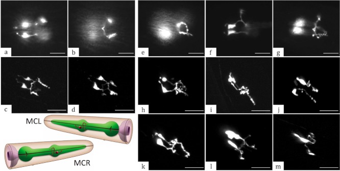
FIG. 6.

The ceh-19 deletion causes axonal defects in MC neurons. In the wild type, MCs have symmetrically located cell bodies and well-organized, consistent axonal projections, as observed by both epifluorescence (a, b) and confocal (c, d) microscopy. In the ceh-19(tm452) mutant background, the ceh-19b::gfp transcriptional reporter fusions were still expressed in the MCs, but revealed various subtle to moderate axonal defects upon epifluorescence (e–g) and confocal microscopy (h–m). All images were captured in the region of anterior pharyngeal bulb with anterior towards the upper left. Strains presented are: UL3012 (a–d), UL3022 (e–g), UL3013 (h–i), UL3019 (j) and UL3022 (k–m). A panel depicting the morphology of MCL and MCR (red) with respect to the pharynx (green) in the wild type is included for comparison (from WormAtlas). Scale bars represent 10 µm in all panels.
