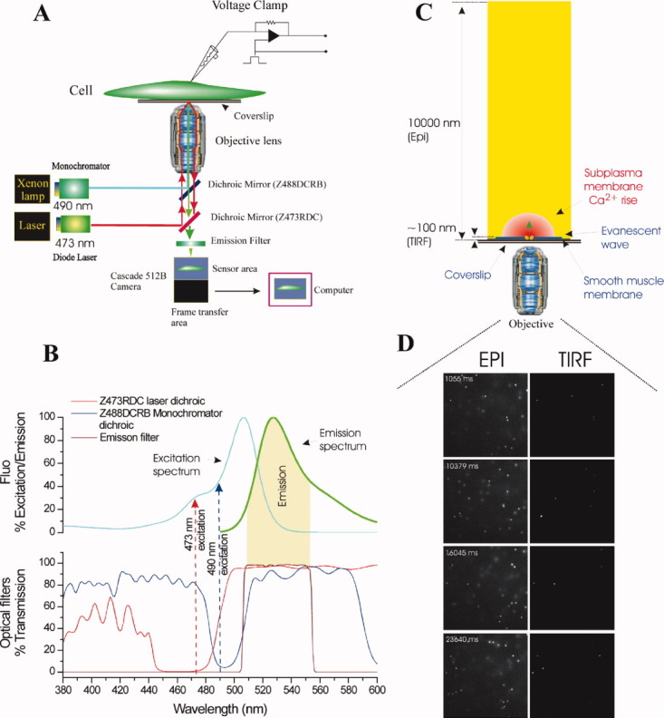
Figure 7.

Schematic of the TIRF and wide-field epi-fluorescence system. (A) The experimental system was constructed around a Nikon TE2000U microscope; excitation and emission light paths are shown. Single cells were voltage clamped in whole cell configuration. Cells were loaded with fluo-5F and illuminated by two separate excitation sources (at 490 nm and 473 nm) via a dual epi-illuminator. The first wavelength (490 nm, bandpass 5 nm; blue), was provided by a monochromator and guided via a fiber optic guide through a field stop diaphragm, a neutral density filter and HQ480/40 excitation filter (not shown) before being reflected off a custom-made long-pass dichroic mirror (Z488DCRB). The latter was transmissive in the ranges 380–480 nm and 506–589 nm and reflective from 485 to 499 nm (B). The second excitation wavelength (473 nm; red), for TIRF illumination, was provided by a blue diode pumped laser and guided via a fiber optic coupling through an iris and 10× beam expander. The 473 nm light was then reflected off a dichroic mirror (Z473RDC) and transmitted through the upper dichroic (Z488DCRB) (A and B) and focused to a spot on the back focal plane of the objective lens (Nikon 60×, oil immersion, NA 1.49). The laser focusing lens was mounted on a micrometer driven translation system so that the laser beam could be adjusted to enter the periphery of the objective aperture to achieve total internal reflection at the interface between the cover glass and the aqueous bathing medium. Fluorescence excited in the specimen by the evanescent wave or wide-field epi-illumination was collected through the objective lens, passed through the dichroic mirrors and a barrier filter and imaged by a Phometrics Cascade 512B or evolve 128 cameras (Roper Scientific). The cameras uses a back illuminated frame transfer CCD with on-chip electron multiplication. (C) Enlarged view illustrating the imaging of near membrane Ca2+ from the microdomain (red) around a single open channel by evanescent wave (blue) formed by the TIRF objective lens and the simultaneously measured wide-field epi-fluorescence depth of field. The depth of the TIRF field was ∼ 100 nm and that of the wide-field epi-fluorescent field ∼ 10000 nm. (D) Single image frames of sub-resolution (100 nm) fluorescent latex beads floating in solution obtained in TIRF (right) and wide-field epi-fluorescence (left). Under wide-field epi-illumination (EPI, D, left) subresolution fluorescent latex beads floating in solution are illuminated through the field. Individual beads drift through Brownian motion. In TIRF illumination (D,TIRF, right) beads are illuminated only when they are within the evanescent field, that is, within ∼ 100 nm of the coverslip. Beads diffusing in and out of the evanescent field appear and disappear suddenly (Reproduced from ref. 45, with permission from Rockefeller University Press).
