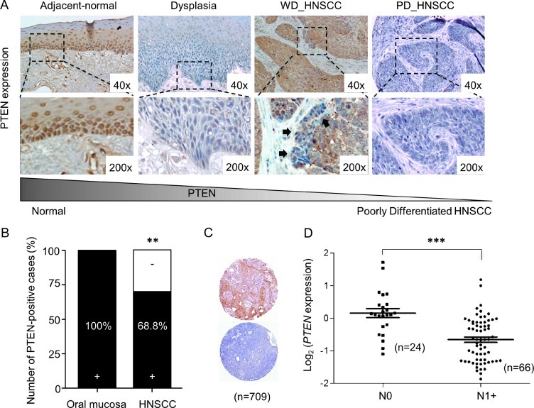
Figure 1.
Loss of PTEN during tumor transformation and lymph node involvement. (A) Representative examples of PTEN expression in normal, dysplastic, and HNSCC from humans demonstrating PTEN expression. Note PTEN down-regulation in the dysplastic epithelium and in the HNSCC epithelial cell invasion (black arrows). (B) Analysis of PTEN expression on oral epithelia (n = 10) and four TMAs for HNSCC (containing n = 709). In normal oral epithelia, PTEN staining was positive mostly in the basal layer. Note an overall PTEN loss in 31.2% of all HNSCC tumors (**P < .01). (C) Representative examples of PTEN staining in HNSCC TMA tumor core (n = 709; positive, top; negative, bottom). (D) Scatter plot of relative microarray signal intensities for PTEN mRNA in HNSCC samples from patients with negative lymph nodes (N0) versus presence of lymph node metastases (N1+; error bar, SEM; **P ≤ .01; ***P ≤ .001).

