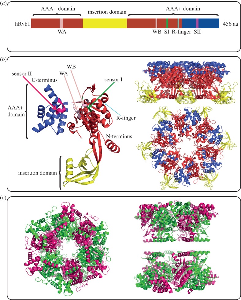
Figure 2.
Overview of the Rvb1/2 structure. (a) Bar graph of the domain organization of human Rvb1. In red is the N-terminal αβα subdomain of the AAA+ domain, in blue is the C-terminal all α subdomain of the AAA+ domain, and in yellow is the insertion domain. Conserved motifs with the AAA+ domain are also highlighted: WA, Walker A; WB, Walker B; SI, Sensor I; SII, sensor II; R-finger, arginine finger. (b) Crystal structure of human Rvb1 monomer on the left-hand side. Side and top view of human Rvb1 hexamer are shown on the right-hand side. The colour scheme used is the same as the one in (a). (c) Top and side views of the crystal structure of dodecameric human Rvb1/2 complex with truncation in part of Domain II. Human Rvb1 monomers are shown in green and human Rvb2 monomers are shown in pink.

