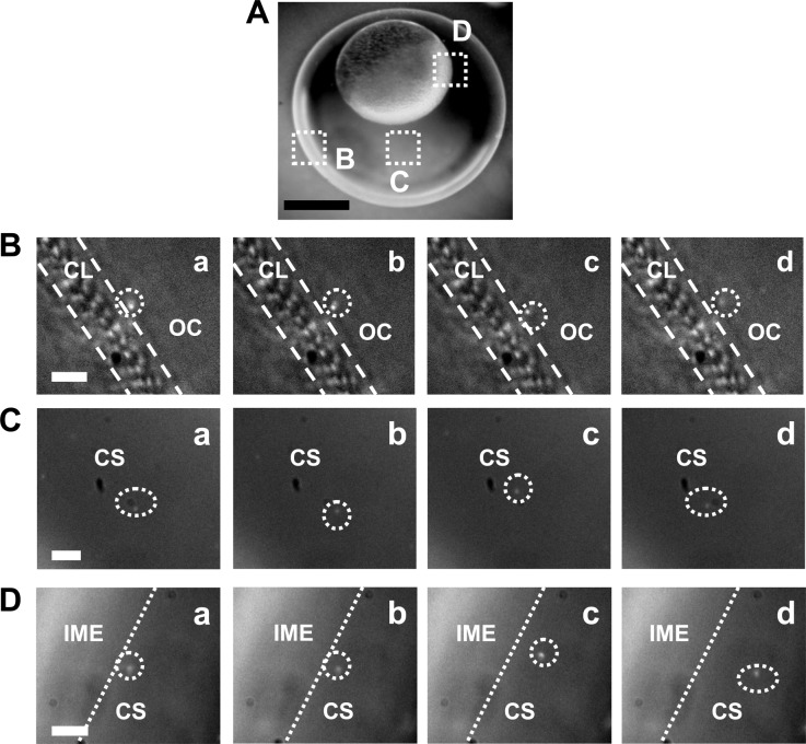
Figure 3.
Real-time imaging of transport of single Au NPs (86.2 ± 10.8 nm; 20 pM) into cleavage-stage zebrafish embryos using DFOMS. (A) Optical image of the cleavage-stage embryo in egg water shows its chorionic layer (CL), chorionic space (CS) and the interface of the CS and the inner mass of the embryo (IME), as highlighted by (B–D), respectively. (B–D) Snapshots of sequential dark-field optical images of the zoom-in areas squared in (A) show the diffusion of single Au NPs (circled) from the egg water (outside chorion, OC) into the CL, in the CS, and at the interface between the CS and IME, respectively. The temporal resolution of real-time imaging is 100 ms. Scale bars are: (A) 200 and (B–D) 5 µm.

