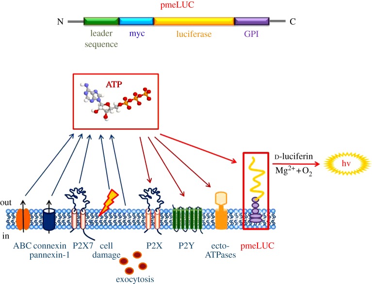
Figure 2.
Membrane topology of pmeLUC. The pmeLUC construct is composed of the full-length coding sequence of luciferase (yellow) inserted in-frame between the N-terminal leader sequence (green) and the C-terminal GPI anchor (violet) of the folate receptor. A c-myc tag (light blue) is also added in-frame for tracking purposes. The pmeLUC protein is targeted and localized to the outer side of the plasma membrane, in close vicinity to all the molecules participating in purinergic signalling (adapted from Pellegatti et al. [24]).

