
Figure 5.
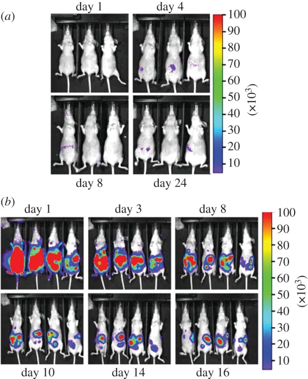
pmeLUC as a probe of ATP within the tumour microenvironment. Bioluminescence imaging of tumour-bearing nude mice injected with HEK293-pmeLUC cells. (a) Healthy nude mice were injected i.p. with 2 × 10⁶ HEK293-pmeLUC cells and monitored for 30 days. No luminescence was detected at any time. (b) Nude/nude mice were injected i.p. with the human ovarian carcinoma cell line OVCAR-3 (1.5 × 10⁶). Twenty days post-inoculum, HEK293-pmeLUC cells (2 × 10⁶) were injected i.p. and luminescence monitored for 16 days. As shown, strong luminescence emission was detected initially throughout the peritoneal cavity, and at later time points at discrete sites corresponding to tumour foci on the peritoneum (adapted from Pellegatti et al. [37]).