Figure 3.

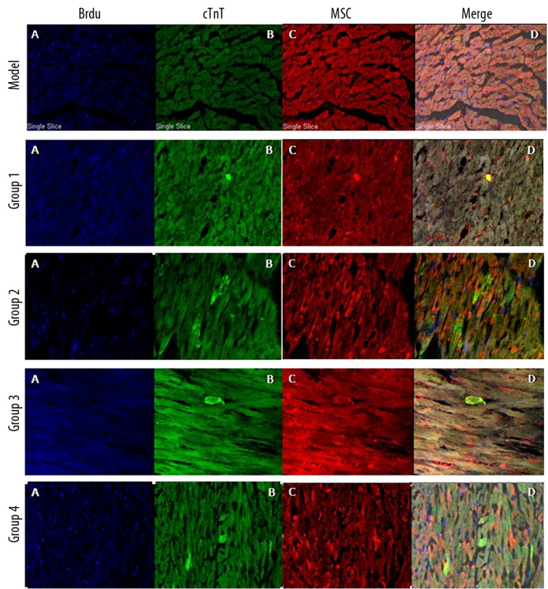
No BrdU-stained cells were present in the model group. BrdU-labelled cells (A) were discretely distributed throughout the myocardial regions of Groups 1, 2, 3 and 4. Some of transplanted BMSCs were also positive for troponin T (B–D).

No BrdU-stained cells were present in the model group. BrdU-labelled cells (A) were discretely distributed throughout the myocardial regions of Groups 1, 2, 3 and 4. Some of transplanted BMSCs were also positive for troponin T (B–D).