Figure 4.

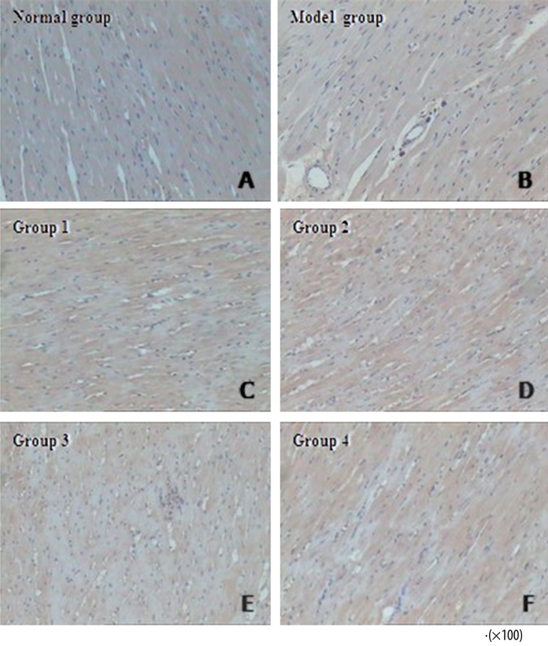
VEGF expression was indicated by brown-yellow staining of tissue sections. VEGF staining was not obvious in the normal group. As compared with the model group, Groups 1–4 exhibited a more robust distribution of VEGF expression.

VEGF expression was indicated by brown-yellow staining of tissue sections. VEGF staining was not obvious in the normal group. As compared with the model group, Groups 1–4 exhibited a more robust distribution of VEGF expression.