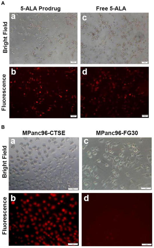
Figure 1. 5-ALA prodrug is efficiently activated by Cath E-expressing PDAC cells.
(A) Representative microscopic images of unfixed human pancreatic cancer cells (MPanc96-CTSE) after treatment with 0.5 μM 5-ALA prodrug (a, and b) free 5-ALA as a positive control (c, d). Cells imaged on an inverted epifluorescence microscope in bright field (top) and TRITC (Tetramethyl Rhodamine isothiocyanate) channel (λex=557 nm, λem=576 nm) (bottom). Images show significant fluorescence signals originating from cells treated with the 5-ALA prodrug, comparable to that obtained with free 5-ALA, indicating the efficient release of 5-ALA from the prodrug. (B) 5-ALA prodrug activation is Cath E-dependent. Representative microscopic images of unfixed PDAC cells expressing Cath E (MPanc96-CTSE) (a, b) and parent cell line (MPanc96-FG30) (c, d) after treatment with 0.5 μM 5-ALA prodrug for 1hr at 37°C. The pronounced fluorescence signal observed within the MPanc96-CTSE cells indicates strong Cath E-mediated release of 5-ALA, which enables cells visualization in the TRITC channel (b). In contrast, MPanc96-FG30 cells, with limited Cath E expression, failed to show an appreciable fluorescence signal, strongly suggesting the lack of free 5-ALA within the cells (d).

