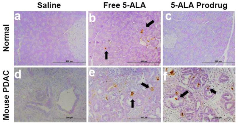
Figure 5. 5-ALA prodrug in combination with light treatment caused selective pancreatic cancer cell death in vivo.

(a–c) Normal pancreas and (d–f) mouse PDAC from genetic mouse model (p53 conditional deletion/LSL-KrasG12D/Pdx1-Cre) treated with 10 J/cm2. (a, d) negative control saline, (b, e) positive control free 5-ALA, and (c, f) 5-ALA prodrug (1 mg 5-ALA equivalent/kg). Formalin-fixed, paraffin-embedded tissue sections were examined for apoptosis by TUNEL using ApopTag Peroxidase Kit. TUNEL staining of PDAC pancreas showed no apoptotic cells in mice treated with saline and PDT (d). However, multiple brown-stained cancer cells were observed in pancreas of animals treated with free 5-ALA and 5-ALA prodrug, indicating apoptosis (e and f, arrows). Tissue sections of normal pancreas of mice treated with PDT using 5-ALA prodrug showed no apoptotic staining, 5-ALA prodrug similar to the negative saline control (a, c). In contrast, scattered brown-stained spots were observed in normal acinar cells in the tissue sections of normal pancreas of mice treated with PDT using free 5-ALA (b, arrows).
