
Fig. 4.
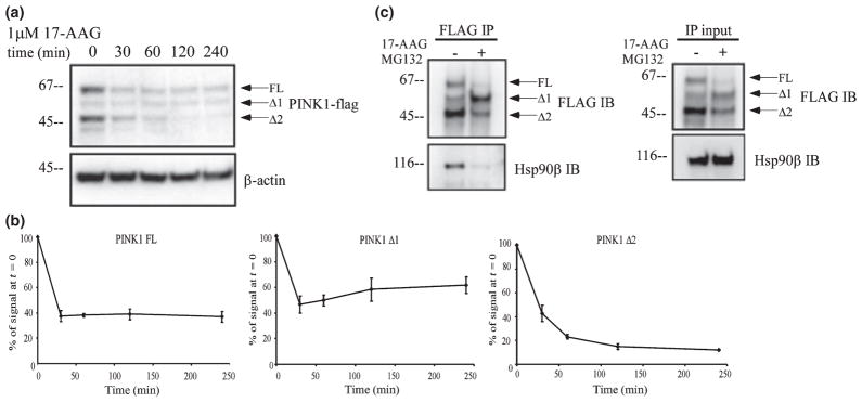
PINK1 interacts with Hsp90. (a) Stably transfected HeLa cells were treated for up to 4 h with 1 μM 17-AAG, an inhibitor of Hsp90 activity. 17-AAG treatment significantly decreased PINK1 Δ2 levels. (b) Quantification of each PINK1 isoform level from three independent 17-AAG experiments as represented in (a). Results are expressed as mean ± SEM. While expression of PINK1 Δ2 continued to decrease over time, level of PINK1 FL and Δ1 did not decrease after 30 min of 17-AAG treatment. (c) FLAG co-immunoprecipitation in HeLa cells transiently transfected with PINK1-flag. Cells were treated with 1 μM 17-AAG for 30 min; 1 μM MG132 was added to prevent 17-AAG-induced degradation. Lysates were immunoprecipitated with anti-flag antibodies and immunoblotted for flag or Hsp90β. Total lysates were used to detect Hsp90β and PINK1-flag level by western blot. 17-AAG led to a dissociation of PINK1-flag from Hsp90β.