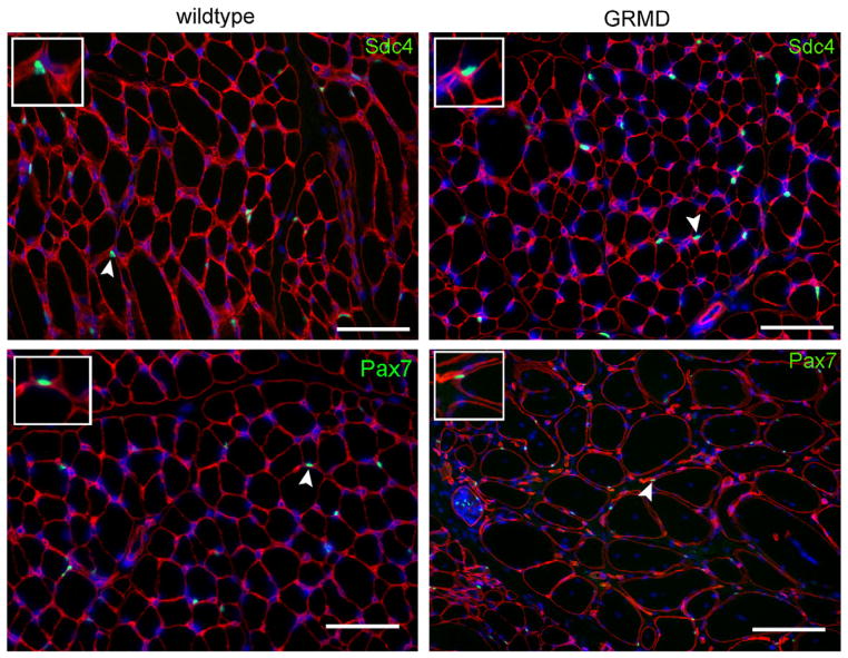
Fig. 3.
Anti-syndecan-4, as well as anti-Pax7, identifies quiescent canine satellite cells in fixed frozen sections. Frozen sections of wild type or GRMD cranial tibial muscle stained for laminin (red) to identify myofiber boundaries. Panels are costained (green) for either syndecan-4 (top row) or Pax-7 (bottom row). Scale bar = 50 μm. Insets: 2× magnified fields containing stained satellite cells indicated by arrowheads.

