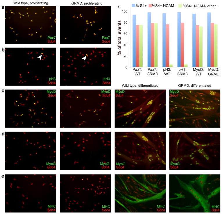
Fig. 4.
Markers of satellite cell identity, proliferation, and differentiation are equivalently distributed in wildtype and GRMD satellite cells in culture. (a) Under proliferation conditions, syndecan-4 and Pax7 are coexpressed by both wildtype and GRMD cells. (b) Under proliferation conditions, mitotic satellite cells identified by anti-syndecan-4 from wildtype and GRMD cultures react with anti-phosphohistone 3 (arrowheads). (c) Under both proliferation and differentiation conditions, satellite cells identified by anti-syndecan-4 from both wildtype and GRMD cultures express MyoD. (d and e) Under proliferation conditions, few syndecan-4 positive satellite cells from either wildtype or GRMD cultures express myogenin or myosin heavy chain, while after three days under differentiation conditions both genotypes display nuclear myogenin and cytoplasmic myosin heavy chain in differentiated myotubes. Myotubes from both genotypes are also qualitatively similar (size, myonuclear number, morphology, etc.). (f) Flow cytometric analysis of mononuclear cells using the immune reagents shown in a–c; distribution of syndecan-4+ve NCAM−ve cells (proliferation-competent myoblasts) is quantitatively similar in all samples. Percentage of proliferation-competent myoblasts that costain for either Pax7, pH3, or MyoD are equivalent in both genotypes. Blue bars, % S4+ (all satellite cell progeny); red bars, % S4+ve NCAM−ve (proliferation-competent myoblasts); green bars, % S4+ve NCAM−veother+ve (proliferation-competent myoblasts positive for Pax7 (first two samples), pH3 (center two samples) or MyoD (last two samples).

