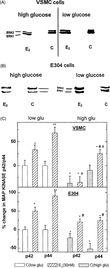
Fig. 2.
(A–C) Western blot depicting the effect of estradiol 17β at 30 nM in the presence or absence of high glucose on p42/p44 MAP-kinase in VSMC (A) and in E304 cells (B). Densitometric analysis of these gels corrected by the concentration of cell protein applied to the gels is shown in (C). Results are means of Western blot analysis of gels from two incubates from three experiments and are expressed as the ratio between the various treatments and control cells at a low glucose. * P < 0.05; ** P < 0.01 for the comparison with control values at low glucose; # P < 0.05 for the comparison with control values at high glucose; and a P < 0.05 for the comparison between the effect of E2 with and without glucose. The statistical analysis was done by ANOVA.

