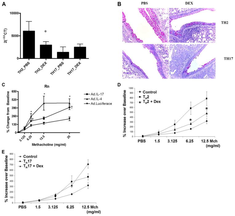
FIGURE 5.
IL-17 is sufficient to induce mucus secretion and AHR. A, gob5 expression was determined in whole lung by quantitative real-time PCR and normalized to the housekeeping gene 18S. Data are graphed as mean ± SEM for n = 4; *, p < 0.05 vs TH2_PBS. B, Expression data were confirmed by PAS staining of lung sections from TH2, and TH17, transferred animals. C, An adenovirus overexpressing IL-4, IL-17, or luciferase control was given i.t. to WT BALB/c (108 PFU/mouse) and airway responsiveness to methacholine was determine on day 3. *, p < 0.05 AdIL-4 vs Adluc; τ, p < 0.5 AdIL-17 vs Adluc. D, Methacholine dose-response curves in control, TH2 vehicle-treated, or TH2 DEX-treated mice. Control mice received all OVA challenges but PBS i.v. at time of cell transfer to experimental groups. Data are graphed as mean ± SEM for n = 6 – 8; *, p < 0.05 for TH2 vs TH2 + DEX. E, Methacholine dose-response curves in control, TH17 vehicle- treated, or TH17 + DEX-treated mice. Control mice received all OVA challenges but PBS i.v. at time of cell transfer to experimental groups. Data are graphed as mean ± SEM for n = 6 – 8.

