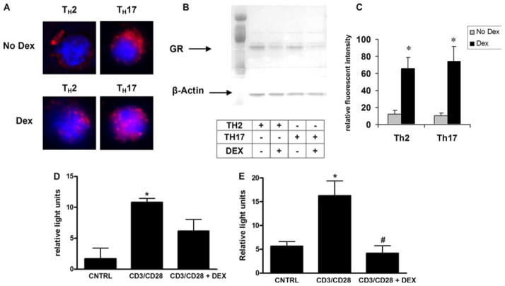
FIGURE 6.
Mechanism of TH17 cell resistance to DEX treatment. TH17 cells are not deficient in their ability to translocate GRα. A, GR staining by immunofluorescent microscopy in DEX-treated (1 μM) and nontreated TH2 and TH17 cells. B, Cytoplasmic protein fractions of DEX-treated and nontreated TH2 and TH17 cells were immunoblotted for GRα and β-actin. Samples were loaded in the following order: TH2_PBS, TH2_DEX, TH17_PBS, and TH17_DEX. C, Florescent intensity of nuclear GRα staining in TH2 and TH17 cells with and without DEX treatment. Data are graphed as mean ± SEM; *, p < 0.05 for DEX compared with no DEX conditions. D and E, TH2 (C) or TH17 (D) cells were transfected with NFAT-luciferase before stimulation with anti-CD3 and anti-CD28 microbeads (6 h) with or without pretreatment (2 h before beads) with DEX; *, p < 0.05 control (CNTRL) vs CD3/CD28; #, p < 0.05 CD3/CD28 vs CD3/CD28 + DEX.

