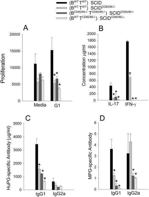
Figure 3.
Decrease in T cell and antibody in SCIDCD80/86−/− recipient mice. (A) T cell proliferation was measured by 3H-thymine incorporation in PG activated spleen cell cultures, (B) cytokine levels in supernatants from PG activated spleen cells, (C) serum human PG-specific antibody response, (D) serum mouse PG-specific. Data represents the mean and SEM (n=7). Data represent the mean and SEM (n=7). * represents statistically significantly differents p<0.05 from the control group (BWTTWT)→ SCID.

