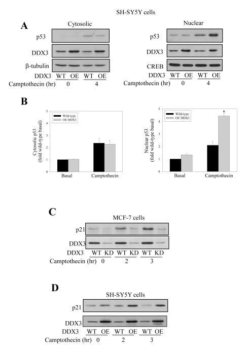
Figure 6. DDX3 stabilizes nuclear p53and increases p21 in response to DNA damage.
(A) Cytosolic (left panels) and nuclear (right panels) fractions were prepared from SH-SY5Y wild-type (WT) cells and SH-SY5Y cells after DDX3 overexpression (OE) by transient adenovirus infection with or without camptothecin (10 μM; 4 hr) treatment, and immunoblotted for p53 and DDX3. β-tubulin and CREB were used as cytosol and nuclear loading controls, respectively. (B) p53 quantitative values are from basal and 4 hr camptothecin-treated cytosolic and nuclear samples in SH-SY5Y wild-type (WT) and DDX3 over-expressing (OE) cells. means ± SEM; n=3; *p<0.05 (C) Wild-type and DDX3 knockdown MCF-7 cells were treated with 10 μM camptothecin for 0, 2 and 3 hr, followed by p21 and DDX3 level measurements. (D) Wild-type SH-SY5Y cells and DDX3-overexpressing SH-SY5Y cells were treated with camptothecin for 0, 2 and 3 hr, and protein levels of p21 and DDX3 were measured. Experiments were repeated three times with similar results.

