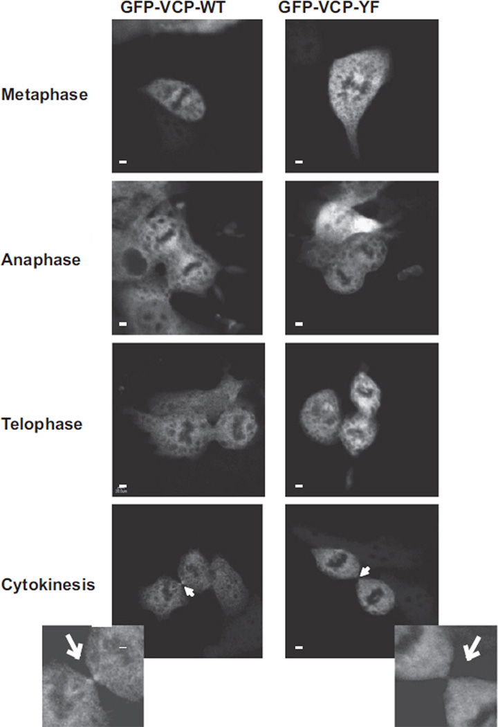
Fig. 8.
VCP is localized to the midbody. HEK293 cells were transiently transfected with GFP-VCP wild-type (WT) or GFP-VCP-Y805F mutant using Fugene-6. Thirty-six hours post-transfection cells were fixed and imaged. Pictures show representatives of cells at cytokinesis from both VCP-WT and VCP-Y805F transfected HEK293 cells, respectively. Cells at cytokinesis were counted for each WT (n = 13) and Y805F (n = 10) and scored for the presence of GFP-VCP at the midbody. The insets are digitally enlarged images of the midbody, marked by the white arrowhead. The scale bars in each panel represent 20 µm.

