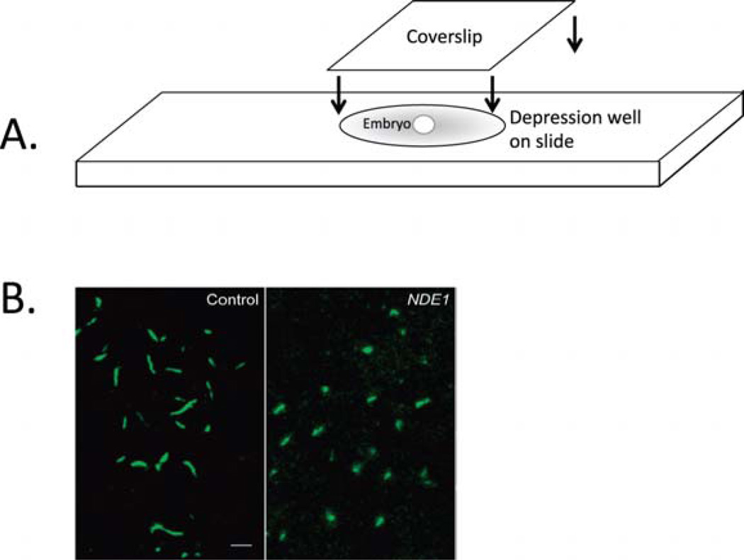
Fig. 1.
Visualization of cilia in whole-mount fixed embryos. (A) After immunostaining for acetylated α-tubulin,whole embryos are mounted in depression well slides and covered with a coverslip. Once placed in the well, the embryo is rotated by gentle rotation of the coverslip until the desired orientation for imaging is achieved. (B) Confocal imaging of cilia in Kupffer’s vesicle in immunostained, mounted embryos. Cilia conformation and length can be easily observed. In this example, control embryos exhibit normal length cilia, whereas NDE1-overexpressing embryos exhibit shorter cilia (modified from Kim et al., 2011). (Permission from Nat. Cell Biol., License #: 2721450638335.) (For color version of this figure, the reader is referred to the web version of this book.)

