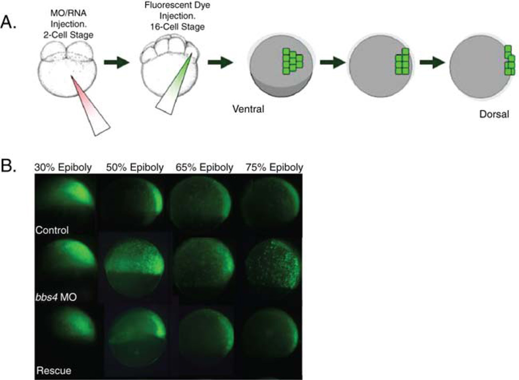
Fig. 4.
Labeling of cells and tracking of movements through gastrulation. (A) After embryo treatment with morpholino or RNA injection at the 1- to 2-cell stage, fluorescent dye is injected into a single marginal blastomere at the 16-cell stage. At gastrulation stages, fluorescing cells can be tracked to monitor movement. (B) Ciliary morphant embryos (bbs4 shown here) exhibit defects in the characteristic movements whereby cells converge on and extend along the dorsal axis. This defect can be rescued by co-injection of wild-type mRNA (modified from Zaghloul et al., 2010). (Permission from Nat. Cell Biol., License #: 2721450638335). (See color plate.)

