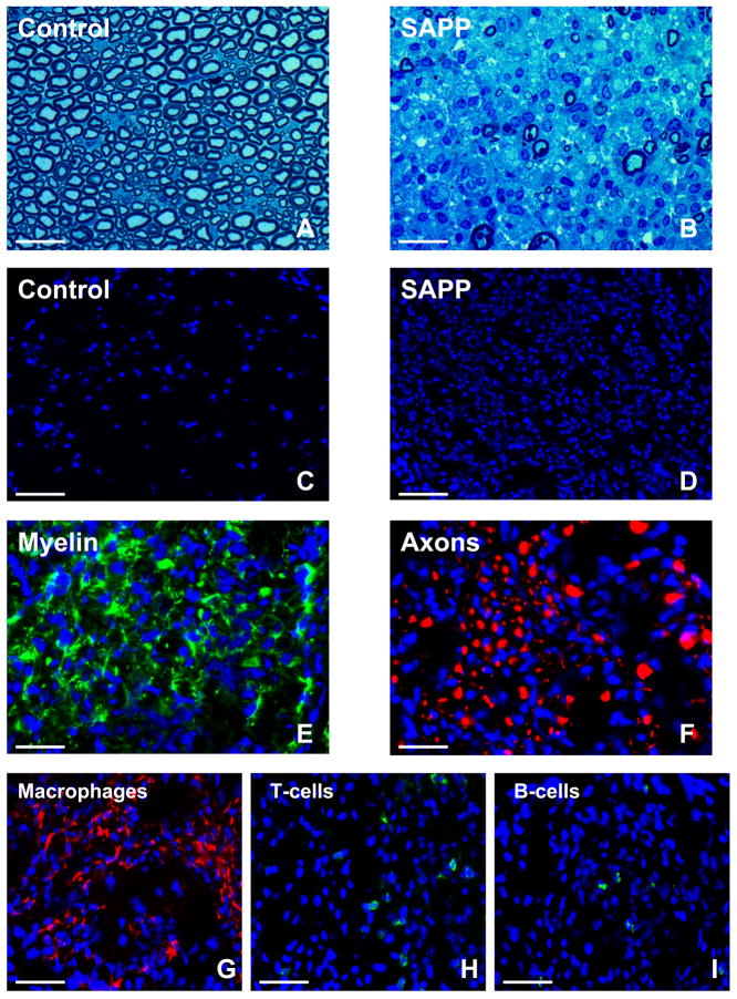
Figure 3. Histopathological features of SAPP.
Representative toluidine-blue photomicrographs of 1 μm glutaraldehyde fixed, osmium tetroxide post-fixed plastic-embedded mouse sciatic nerve axial sections show the normal distribution of myelinated axons in control B7-2 deficient non-obese diabetic mice (A) in contrast to the severe demyelination and reduction in axonal density coupled with mononuclear cell infiltration observed in SAPP at 30 weeks of age (B). The extent of intense mononuclear cell infiltration in SAPP is depicted by the DAPI-stained photomicrographs of 10 μm frozen sciatic nerve axial sections (D) compared to unaffected controls (C). SAPP is associated with demyelination, as demonstrated by the loss of the normal honeycomb Schwann cell S100β (green) immunoreactivity (E), and axonal loss/ fragmentation depicted by reduced density and spotty/ filamentous neurofilament-H (red) immunoreactivity (F) as described in severe cases of human CIDP. Mononuclear leukocyte infiltration consists predominantly of F4/80+ macrophages (red immunoreactivity, G). Fewer T-cell (H) and B-cell (I) infiltrates are seen, depicted by green immunoreactivity. Blue depicts nuclei (DAPI stain). Kindly refer to [36] for a detailed histopathological and electrophysiological characterization of SAPP. Magnification bars: A, B, E, F = 25 μm, C, D = 75 μm, G, H, I = 35 μm.

