Figure 5.

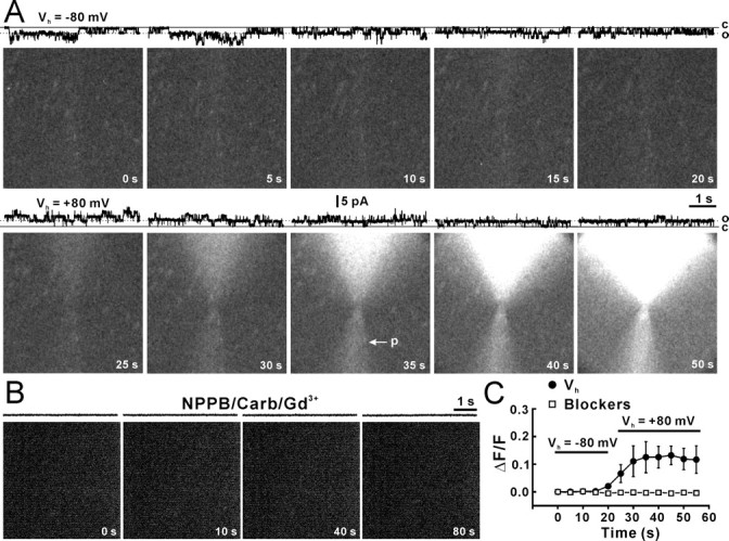
Blockade of ATP influx by negative holding or hemichannel blockers. A, Light induced by ATP influx when an inside-out patch was held at −80 mV (Vh = −80 mV) and +80 mV (Vh = +80 mV). Recording traces at the top of each panel illustrate hemichannel activities at open (o) and closed (c) states. The arrow (p) indicates the pipette containing the enzyme that delivered by a light, but sustained pressure. B, ATP influx when gap-junction blockers, Gd3+ (30 μm), carbenoxolone (Carb, 100 μm), and NPPB (100 mm) were applied in the bath solution. Patches showing hemichannel activity before action of blockers (control) were chosen to move to the enzyme pipette. The inside-out patch was held at +80 mV. C, Relative light intensity (ΔF/F) against time during holding at −80 mV (filled circles, Vh = −80 mV) and +80 mV (filled circles, Vh = +80 mV; n = 7 patches) or in the presence of gap-junction blockers (open squares; n = 9 patches; two-way ANOVA, p < 0.01 compared with absence of blockers). Error bars indicate SEM.
