
Figure 1.
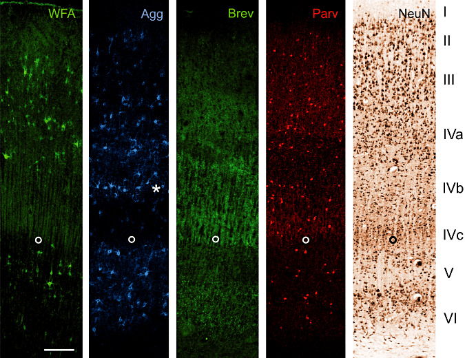
A17 Co: Laminar distribution of major extracellular matrix components in area 17 of human control brain. Two neuronal markers [neuronal nuclear protein (NeuN) and parvalbumin (Parv)] are shown to indicate the basic laminar profile. Wisteria floribunda lectin (WFA) as established marker for perineuronal nets indicates many nets in layer III, IVa and V. Only few perineuronal nets can be seen in layer VI whereas layer I, II and IVc are virtually devoid of WFA‐stained nets. Compared with WFA staining, pan‐aggrecan antibody HAG (Agg) reveals more perineuronal nets, indicating variations in aggrecan glycosylation. This becomes clear especially in layer IVb (asterisk) containing many Agg positive nets but devoid of WFA‐stained perineuronal nets. Brevican antibody B50 (Brev) reveals a clearly different laminar pattern. Immunoreactivity is mainly restricted to layers IVa, IVc and VI. Further, brevican antibody B50 (Brev) does not show the typical perineuronal net‐like appearance but reveals dot‐like structures. The open circle marks the lower edge of layer IVc. Scale bar = 200 µm.