
Figure 5.
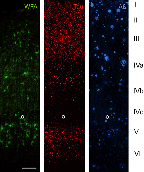
A17 AD: Laminar distribution of perineuronal nets (PNs) detected by Wisteria floribunda lectin (WFA) in area 17 of Alzheimer's disease (AD) brain. Two key pathology markers, hyperphosphorylated tau (Tau) and amyloid beta (Aβ) are shown to demonstrate the basic laminar pathology profile. WFA labels N‐acetylgalactosamine containing PNs in layer III, IVa and V. Only few PNs can be seen in layer VI whereas layer I, II and IVc are virtually devoid of WFA‐stained nets. This laminar distribution resembles the distribution pattern in control brain. Tau pathology to some extent overlaps with PN‐rich layers but PN‐ensheathed neurons invariably are devoid of tau pathology, whereas no obvious relation of amyloid plaque distribution and presence of PNs is detectable. The open circle marks the lower edge of layer IVc. Scale bar = 200 µm.