
Figure 8.
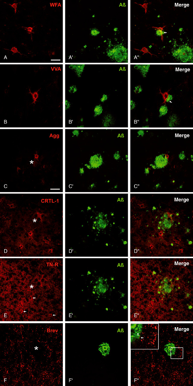
Major extracellular matrix components in spatial relation to amyloid plaque pathology. (A,B) Well‐preserved N‐acetylgalactosamin reactive perineuronal nets (PNs) are located even at the outer zone of the amyloid plaque corona. Occasionally PN‐ensheathed dendrites are penetrating the amyloid deposits (arrows). C. Aggrecan (Agg) core protein immunoreactive nets and D. PNs immunoreactive for link protein 1 occur in close contact to the amyloid plaque territory. E. Tenascin‐R (TN‐R) displays an ubiquitous neuropil immunoreaction and some PNs (arrows) can be detected without obvious alteration in the close proximity of multiple plaques. F. Brevican (Brev) immunoreactive perisynaptic axonal coats of extracellular matrix are clearly stained without evident alterations in the surrounding plaque territory. (Insert in F) Additionally, some axonal coats are even detectable within the plaque corona (arrows) whereas the plaque core is devoid of brevican immunoreactivity. Scale bar in A = 50 µm applies for all pictures.