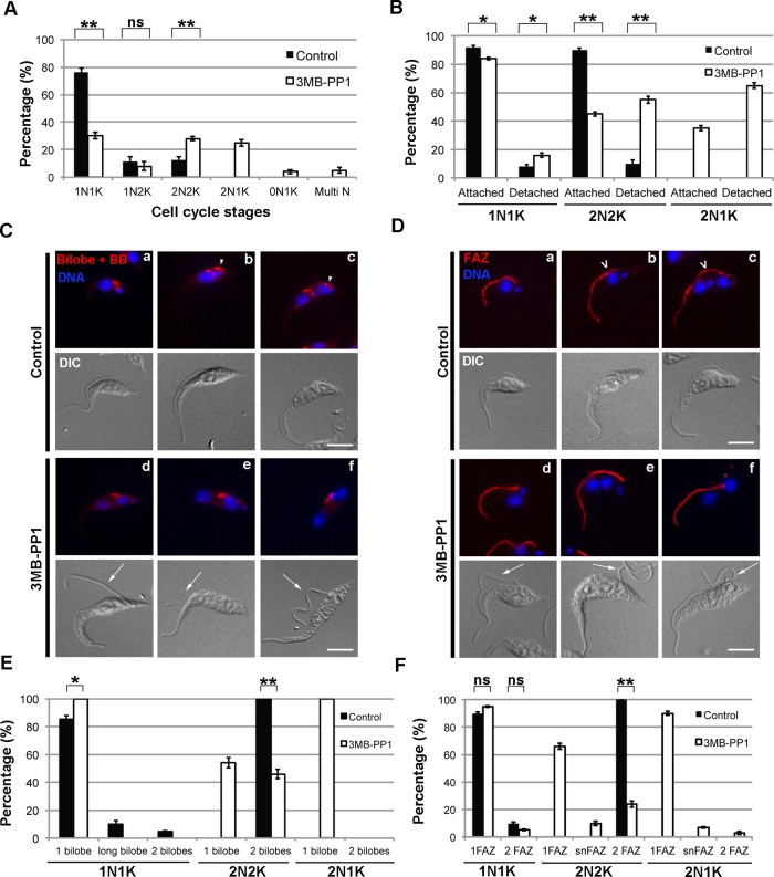
FIGURE 2:
Cells expressing TbPLKas and treated with 3MB-PP1 have DNA, bilobe, and FAZ defects. (A) TbPLKas cells were treated with 3MB-PP1 or vehicle control for 9 h and then fixed and stained with DAPI to label DNA. The DNA content of the cells was determined by fluorescence microscopy. Cells treated with 3MB-PP1 showed an increase in 2N2K cells and the appearance of aberrant 2N1K cells. (B) TbPLKas cells treated as in A were fixed and evaluated for flagellar attachment by differential interference contrast microscopy. Cells treated with 3MB-PP1 showed high levels of flagellar detachment. (C) TbPLKas cells were treated as in A and then fixed and labeled with anti-TbCentrin4 antibody (Bilobe + BB; red) and DAPI (DNA; blue) to label DNA. The bilobe is present between the kinetoplast and nucleus (a). The new bilobe forms toward the posterior of the cell (arrowhead) in a subset of 1N1K cells (b) and moves away from the old bilobe as the cell cycle progresses (c). Cells treated with 3MB-PP1 had difficulty duplicating the bilobe (d–f) and have detached new flagella (arrows). (D) TbPLKas cells were treated as in A and then fixed and labeled with anti-FAZ1 antibody (FAZ; red) and DAPI (DNA; blue) to label DNA. The FAZ underlies the flagellum and runs from the posterior to the anterior end of the cell (a). The new FAZ (empty arrowhead) forms toward the posterior of the cell (b), then extends until just before cytokinesis (c). Cells treated with 3MB-PP1 were not able to assemble a new FAZ and had detached flagella (d–f; arrows). (E) Quantitation of the cells in C. (F) Quantitation of the cells in D. Scale bars, 5 μm. Error bars, SD of three biological replicates with 300 cells counted per condition. *p < 0.05, **p < 0.01, ns, not significant.

