FIGURE 3:
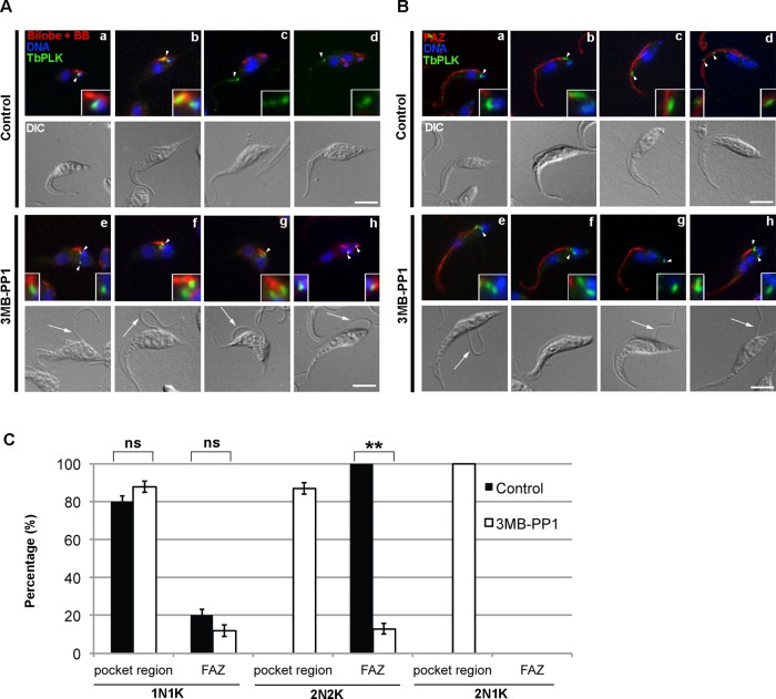
Inhibition of TbPLK causes the kinase to remain in the pocket region. TbPLKas cells were treated with 3MB-PP1 or vehicle control for 9 h, then fixed and stained with anti-TbPLK antibodies and organelle markers to identify the location of TbPLK. Insets in A and B are threefold magnifications of the area around the TbPLK signal. (A) Cells were stained with anti-TbCentrin4 (Bilobe + BB; red), anti-TbPLK (TbPLK; green), and DAPI (DNA; blue). In control cells TbPLK is initially present in the pocket region, which consists of the basal body, MtQ, and bilobe (a, b; arrowheads), then migrates out toward the anterior of the cell (c, d; arrowheads) once kinetoplast and nuclear duplication occur. In 3MB-PP1–treated cells, the kinase remains in the pocket region even after nuclear duplication had occurred (e–h; arrowheads). (B) Cells were stained with anti-FAZ1 (FAZ; red), anti-TbPLK (TbPLK; green), and DAPI (DNA; blue). In the control cells the TbPLK signal shows the same migration pattern as in A (a–d; arrowheads). In 3MB-PP1–treated cells, the kinase does not migrate toward the posterior of the cell (e–h; arrowheads) and remains near the flagellar pocket. (C) Quantitation of the data in A and B. Scale bars, 5 μm. Error bars, SD of three biological replicates with 300 cells counted per condition. **p < 0.01, ns, not significant.

