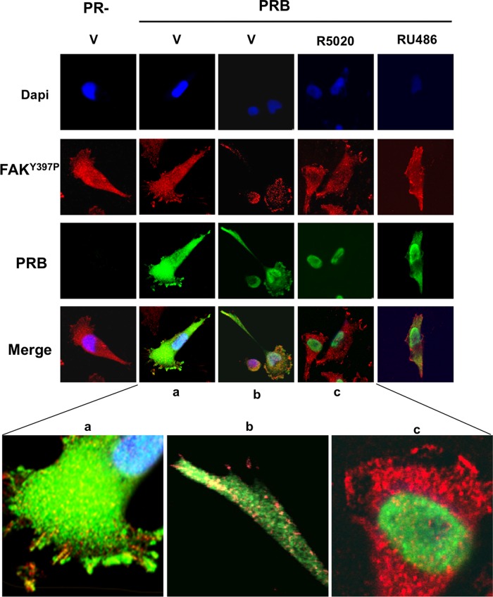
FIGURE 5:
PRB and FAKY397p are colocalized in focal adhesion sites. MDA-iPRAB cells were induced (PRB) or not (PR−) by Dox for 24 h and then treated with either 10−8 M R5020 or 10−6 M RU486 or vehicle for 30 min. Immunofluorescence microscopy was performed as described in Materials and Methods by analyzing FAKY397P (red) and PRB (green). The nuclei were counterstained with DAPI (blue). Photographs were taken by using a confocal microscope at 400× magnification. (a–c) Magnifications to focus on representative structures.

