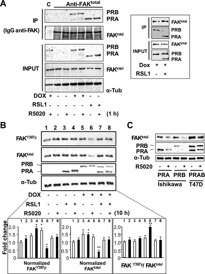
FIGURE 6:
PRA and PRB interact with FAK complexes and regulate their turnover. (A) MDA-iPRAB cells were induced by RSL1 and/or Dox for 24 h and then exposed to 10–8 M R5020 or vehicle for 1 h. Cell lysates were incubated with either total FAK antibody or nonrelated antibody (immunoglobulin G). Lysates (input) and immunoprecipitates (IPs) were analyzed by Western blot for FAKY397P, FAKtotal, PRA, PRB, and tubulin. Framed inset: the coIP experiments were repeated using iPRAB cells induced by either Dox or RSL1 + Dox to induce PRB or PRA + PRB for 24 h. (B) iPRAB cells were induced by either RSL1 or Dox or both of them and then treated or not by 10−8 M R5020 for 10 h. Western blots were performed and quantified as described in Figure 4A for FAKY397P, FAKtotal, PRA, PRB, and tubulin (mean ± SEM, Mann–Whitney statistical test). (C) Ishikawa cells stably transfected by either PRA or PRB and T47D cells endogenously expressing PRA and PRB were treated either by 10−8 M R5020 or vehicle for 24 h. Western blot analyses were performed as in B.

