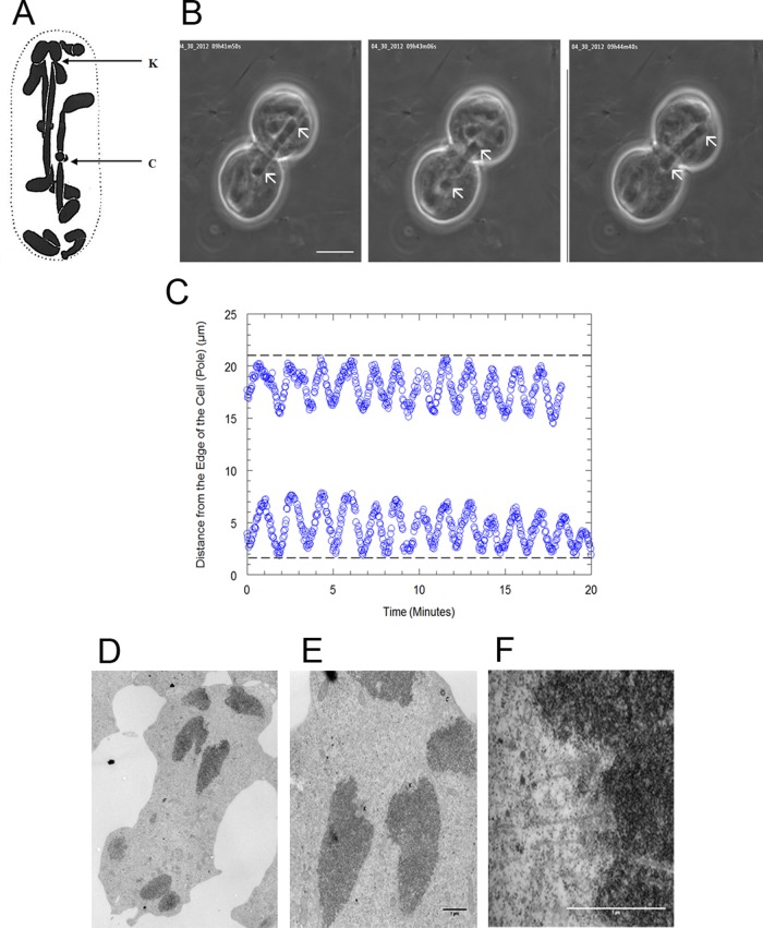
Figure 1:
(A) Fixed and sectioned Mesostoma spermatocyte taken from Husted and Ruebush (1940), showing three bivalents and four univalents. The arrow labeled K points to the kinetochore of a bivalent, and the arrow labeled C points to a chiasma. (B) Montage of phase contrast microscope images of a Mesostoma spermatocyte, illustrating a bivalent as it moves to and away from the spindle poles during prometaphase/metaphase. The arrows indicate the position of the kinetochores. Mesostoma spermatocytes have a precocious cleavage furrow, which begins ingression when bivalents achieve bipolar orientation in prometaphase and then stalls, giving the spermatocytes a dumbbell-shaped appearance (Forer and Pickett-Heaps, 2010). Bar, 10 μm. (C) Distance of the kinetochores of partner half-bivalents from the edge of the cell (pole) in micrometers vs. time in minutes in an M. ehrenbergii spermatocyte. In this cell the average away-from-pole velocity is 6.9 μm/min and the average to-the-pole velocity is 7.5 μm/min. (D–F). Electron microscopy images of a Mesostoma spermatocyte. (D) A low-magnification overview image of a Mesostoma spermatocyte, illustrating two half-bivalents and two univalents at the upper pole. (E) Higher-magnification image of D illustrating the two kinetochores (K) of two half-bivalents and the centriole (C), which is embedded in the pericentriolar material. (F) Higher-magnification image of the kinetochore (K) of the right half-bivalent from E, illustrating microtubules terminating at the kinetochore. Bar, 1 μm.

