
Figure 5:
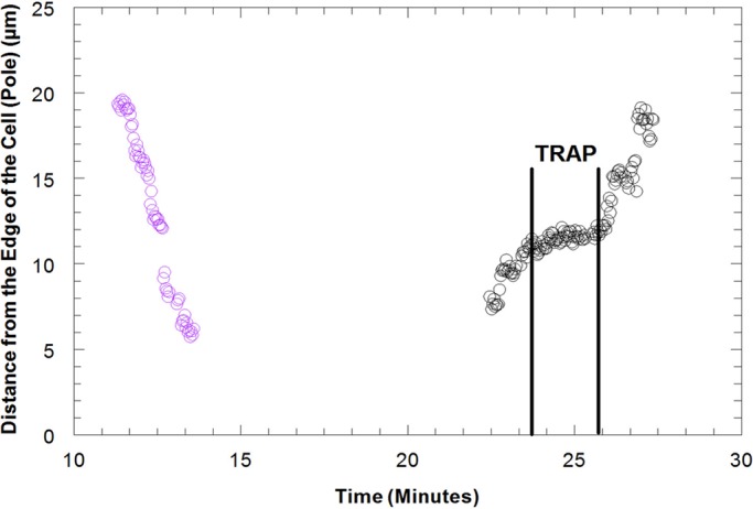
Distance of the kinetochore of a univalent from the edge of the cell (pole) in micrometers vs. time in minutes as the univalent moves from the upper spindle pole to the lower spindle pole and then from the lower spindle pole back to the upper spindle pole in a Mesostoma spermatocyte. The univalent (purple circles) moved from the upper pole to the lower pole with a velocity of ∼7.0 μm/min. A power of 15 mW in the trap, illustrated by the two vertical lines, was applied as the univalent reoriented and segregated from the lower pole back to the upper pole. The trap caused the univalent to decrease in velocity to 0.33 μm/min and then stop. When the trap was released, the univalent moved to the upper pole with its original velocity.