FIGURE 1:
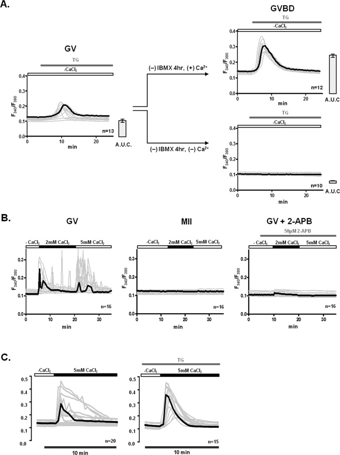
[Ca2+]e and Ca2+ influx are required to fill [Ca2+]ER in oocytes. The underlying Ca2+ influx mechanism(s) are inactivated during maturation and are sensitive to 2-APB and TG at the GV stage. (A) The contribution of extracellular Ca2+ to [Ca2+]ER content was estimated in GVBD-stage oocytes after culturing GV oocytes for 4 h in media supplemented with 1.7 mM CaCl2 or without supplementation, nominal Ca2+-free medium. Release of [Ca2+]ER was induced by addition of 10 μM TG. All [Ca2+]i responses are shown in the graphs, and the bold trace in each graph represents the mean response; bar graphs to the right of each Ca2+ panel denote mean ± SEM of [Ca2+]ER content estimated as area under the curve. (B) Spontaneous Ca2+ influx was measured in GV oocytes and MII eggs. Oocytes and eggs were placed in Ca2+-free conditions, after which 2 and 5 mM CaCl2 were successively added. Given that only GV oocytes showed Ca2+ influx, they were pretreated with 50 μM 2-APB for 5 min before addition of CaCl2 to prevent influx. (C) Ca2+ influx was promoted by addition of CaCl2 into GV oocytes with and without prior treatment with TG. GV oocytes were placed in nominal Ca2+-free media or exposed to 10 μM TG for 30 min in nominal Ca2+-free medium to deplete [Ca2+]ER, after which 5 mM CaCl2 was added. Representative traces are shown, and bold trace represents mean response.

