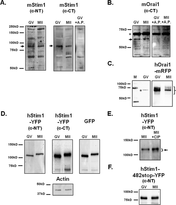
FIGURE 3:
Mouse oocytes express Stim1 and Orai1. (A, B) The expression of Stim1 and Orai1 molecules was probed using lysates of GV oocytes (n = 100 and 200, respectively) and MII (n = 100 and 200, respectively) eggs and specific antibodies. (A) Left and middle, black arrows point to the band corresponding to Stim1. An antigenic peptide was used to confirm the specificity of the anti–C-terminus Stim1 antibody. (B) Right, black arrow points to the band that represents Orai1. The upper band of ∼68 kDa marked with an asterisk is believed to be nonspecific reactivity, as it was not abolished by pretreatment with the antigenic peptide. (C) hOrai1-mRFP expression (n = 16 GV oocytes/MII eggs) was analyzed in overexpressing cells using the same antibody. (D) Heterologous expression of hStim1-YFP was demonstrated in lysates of oocytes/eggs (n = 45) using the anti–N-terminus and anti–C-terminus Stim1 antibodies and an anti-GFP antibody. The actin protein was probed as a loading control (bottom, middle). (E) To examine the possible phosphorylation of hStim1-YFP in mouse oocytes, lysates of hStim1-YFP–expressing MII oocytes (n = 18) were incubated with or without calf intestine phosphatase and then immunoblotted with anti-Stim1 antibody (right). The presence of hStim1 is denoted by an arrow, and the bracket denotes the presence of polypeptides of different MWs. (F) hStim1–482-stop-YFP–overexpressing GV and MII cells (n = 18) were probed with the anti–N-terminus Stim1 antibody, and blots are shown.

