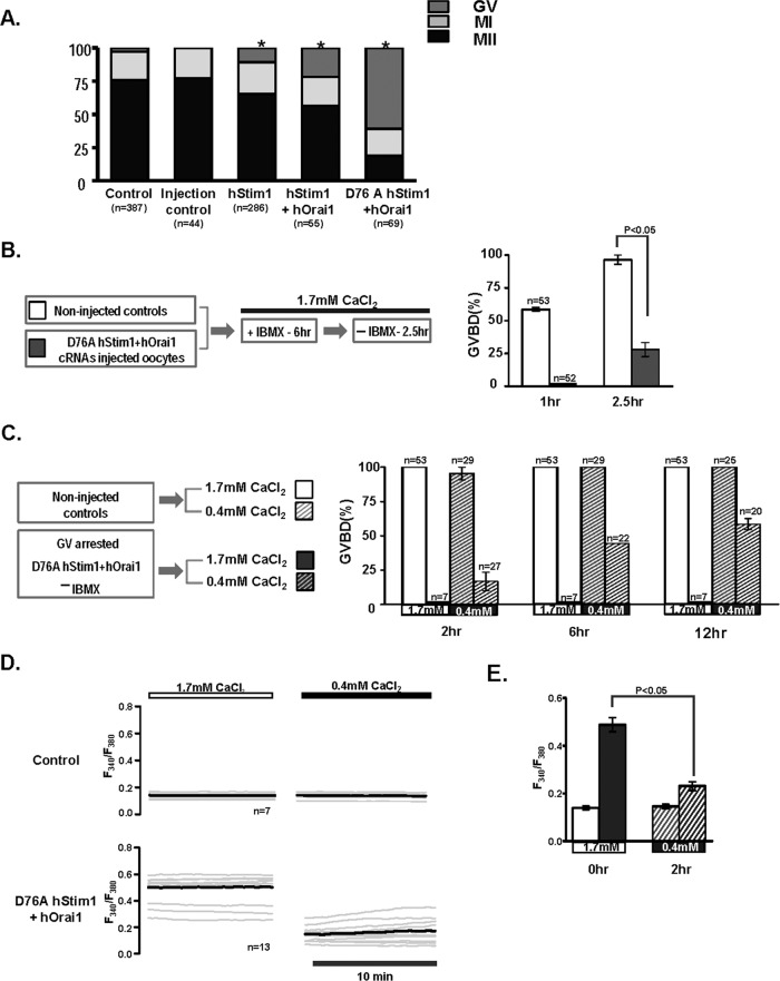
FIGURE 7:
Changes in Ca2+ homeostasis affect resumption of meiosis and oocyte maturation. (A) Control, sham-injected, and oocytes expressing hStim1-YFP, hStim1-YFP+hOrai1-mRFP, or D76A Stim1-YFP+hOrai1-mRFP were in vitro matured and their maturation rates assessed. Expression of any mRNAs that increased basal [Ca2+]i even transiently caused a reduction in maturation rates, although expression D76A hStim1-YFP+hOrai1-mRFP nearly completely prevented resumption of meiosis. Asterisks above bars represent treatments that significantly reduced GVBD rates (p < 0.05). (B) The effect of coexpression of D76A hStim1-YFP + hOrai1-mRFP on meiotic resumption was investigated as depicted in the flow chart (left), and data are summarized in the bar graph (right). Expression of D76A hStim1-YFP+hOrai1-mRFP in GV oocytes delayed and mostly prevented GVBD under normal [Ca2+]e (p < 0.05). (C) As before, but selected GV-arrested oocytes were transferred to low-[Ca2+]e medium (0.4 mM), which partly rescued the arrest caused by expression of D76A hStim1-YFP+hOrai1-mRFP. (D) Ca2+ traces depicting the effect of lowering [Ca2+]e on basal [Ca2+]i in control and GV-arrested D76A hStim1+hOrai1–expressing oocytes monitored before and 2 h after lowering [Ca2+]e from 1.7 to 0.4 mM; the bold trace represents the mean of the responses. (E) A bar graph was used to summarize in the same group of oocytes the mean ± SEM change in basal [Ca2+]i caused by lowering external [Ca2+]e; p < 0.05.

