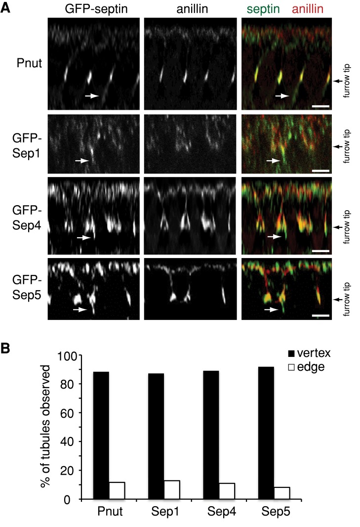
FIGURE 3:
All septins localize to CFT-tubules at the vertices of intersecting cleavage furrows. (A) The z-sections of embryos expressing Pnut-GFP, Sep1-GFP, Sep4-GFP, or Sep5-GFP and immunostained with an anti-anillin antibody to mark the furrow tip. Bar, 5 μm. (B) Quantification of the localization of tubules positive for different septin-GFP fusion proteins relative to the vertex of intersecting cleavage furrows. The data are the combined number of tubules observed in at least 10 embryos for each condition.

