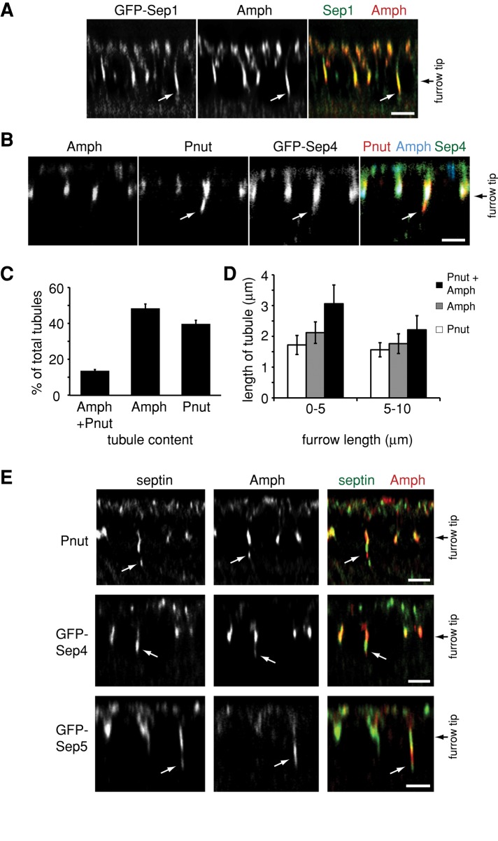
FIGURE 4:
Different subpopulations of CFT-tubules are defined by different septin complexes. (A) A z-section of an embryo from a Sep1-GFP–expressing Drosophila line immunostained with an anti-amphiphysin (Amph) antibody. White arrows point to CFT-tubules. (B) A z-section of an embryo from a Sep4-GFP–expressing Drosophila line immunostained with an anti-amphiphysin (Amph) antibody and an anti-Pnut antibody. White arrows point to CFT-tubules. (C) Quantification of the number of CFT-tubules that stain uniquely with Pnut or amphiphysin (Amph) and CFT-tubules that possess both markers. Error bars, ±SD. n = 11 embryos. (D) Quantification of the length of different CFT-tubule populations at different stages of cleavage furrow ingression. Error bars, ±SD. n = 11 embryos. (E) Different septin-GFP–expressing embryos stained with an anti-amphiphysin (Amph) antibody that define CFT-tubules that have different septin and amphiphysin subdomains. White arrows point to CFT-tubules. Bar, 5 μm.

