
Figure 3.
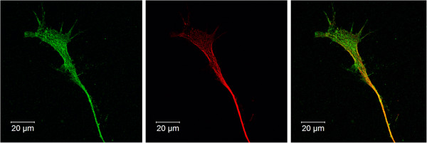
Growth cone organization 120 h after RA treatment. M17 neuroblastoma cells were grown on cover slips. Cells were fixed, stained, and immunofluorescent images were taken (63X). Synapsin-1/2 (green), β3-tubulin (red) and nuclei (blue). β3-tubulin expression is predominantly localized to the neurite body, whereas synapsin-1/2 accumulates within the growth cone.