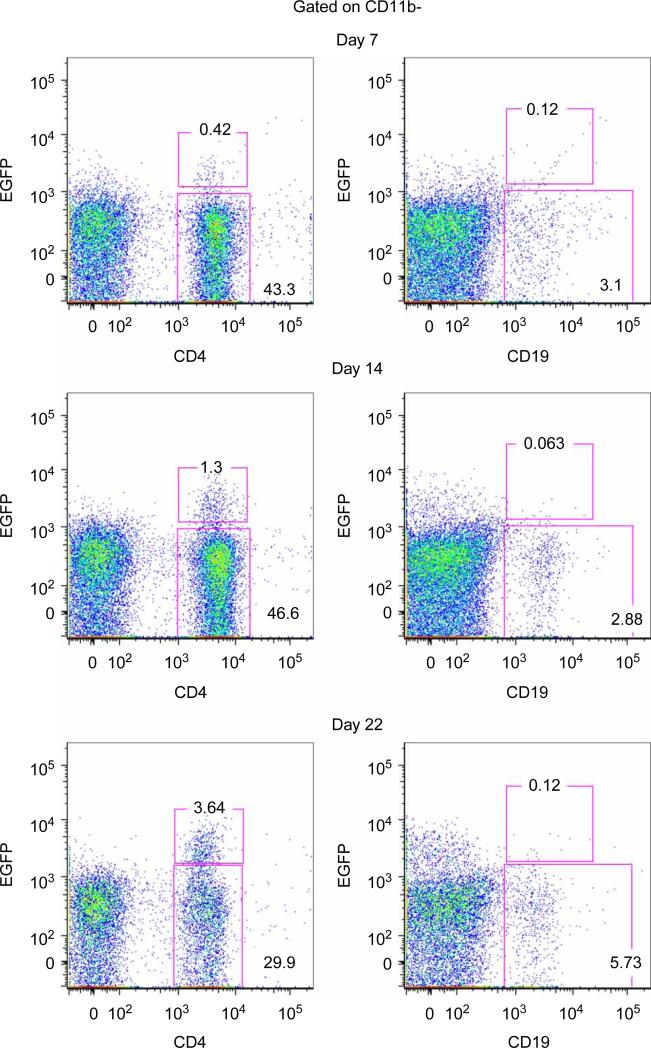
Figure 2.
CD4+ cells produce IL-10 in the CNS during EAE. EAE was induced in IL-10–IRES–eGFP reporter mice by adoptive transfer of 1 × 10⁶ encephalitogenic T cells per mouse. Clinical signs of EAE were scored daily. At onset (day 7), peak (day 14) and resolution (day 22) of EAE disease the percentages of CD11b–-gated CNS infiltrating CD4+ and CD19+ that were EGFP– and EGFP+ were determined by flow cytometry and the percentage of each population is indicated on the graphs. Representative data from one of three mice at each time point is shown.

