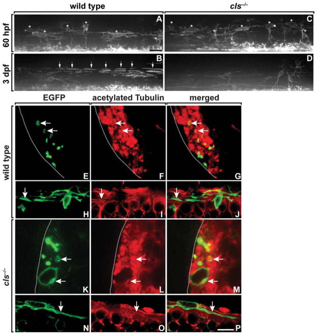
Fig. 3.
OPCs initiate axon wrapping in cls mutant embryos. A–D: Lateral views of spinal cords, dorsal up, of wild-type and cls−/− embryos and larvae carrying the Tg(nkx2.2a:megfp) reporter to mark the myelinating subset of oligodendrocyte lineage cells. Asterisks mark dorsally migrated OPCs and arrows indicate axon wrapping. OPC number and morphology are similar in wild-type and cls−/− embryos at 60 hpf (A, C), but mutants have a deficit of oligodendrocytes at 3 dpf (B, D). Transverse (E–G, K–M) and sagital (H–J, N–P) sections of 64 hpf wild-type and cls−/− spinal cords expressing the Tg(nkx2.2a:megfp) reporter (green) and labeled with anti-acetylated Tubulin (red). OPC processes ensheath axons in both wild-type (E–J) and mutant (K–P) embryos. Scale bars, 24 μM (A–D), 4.5 μM (E–G, K–M), and 9 μM (H–J, N–P).

