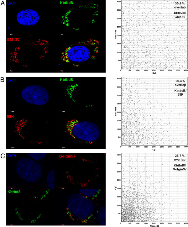
Figure 5.
Co-localization of KBTBD8 and GM130, 58 K and Golgin97. BJ cells were fixed and co-stained with KBTBD8 antibody and the Golgi markers GM130 (A), 58 K (B) or Golgin97 (C). Overlapping of the two fluorescence pictures was analyzed using the Fluoview Software driving the CONFOCAL LASER SCANNING BIOLOGICAL MICROSCOPE (Olympus). A: Co-staining of KBTBD8 and GM130 showed a 34.4% co-localisation. B: Co-staining of KBTBD8 and 58 K showed a 20.4% co-localisation. C: Co-staining of KBTBD8 and Golgin97 showed a 28.7% co-localisation. Scale bars as indicated.

