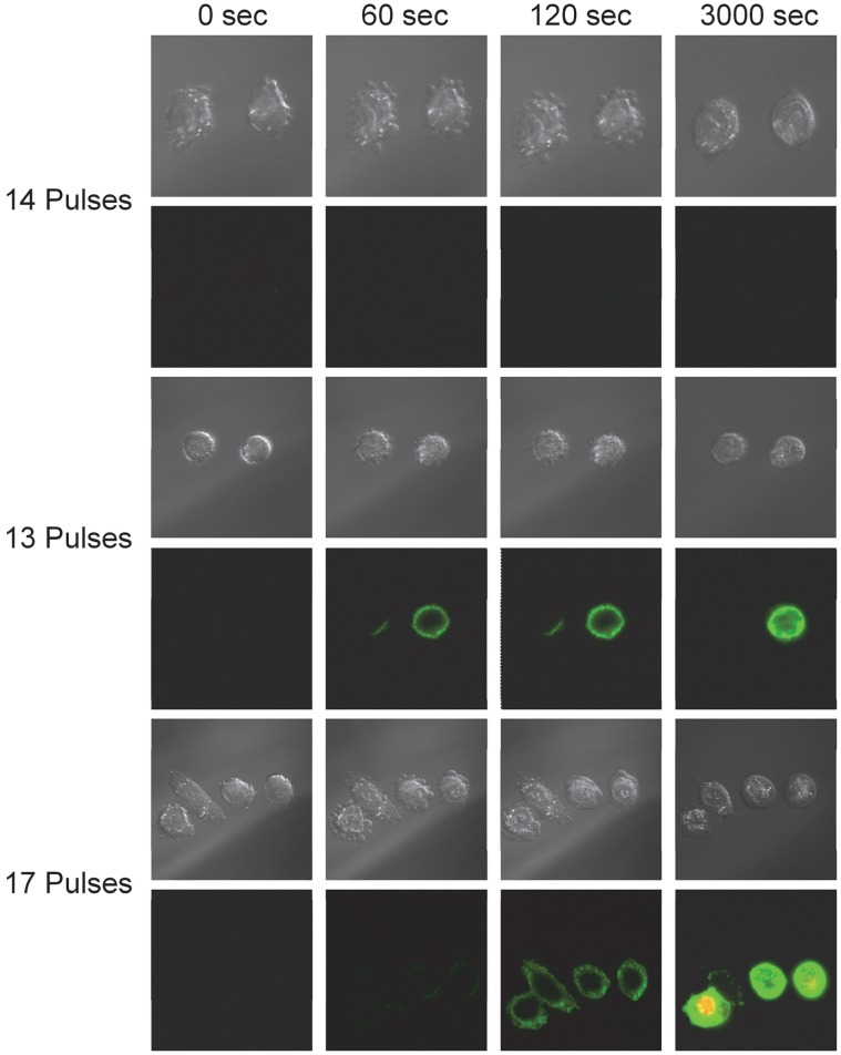
Figure 3. Representative images of the diverse reaction of CHO-K1 cells to nsPEF exposure.
Series 1: Cells undergoing morphological changes without the observation of PS externalization. Series 2: Two cells both expressing PS acutely, but one cell recovers while an adjacent cells remains expressive at 50 minutes post exposure. Series 3: Four cells exposed to the same conditions, all indicate PS expression, but only one cell expresses PS initially and overtime uptakes PI into the interior of the cell.

