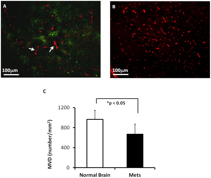
Figure 6. Immunohistochemical study of microvascular density (MVD) in brain metastases.
A. Anti-CD31 staining was performed on a brain section bearing metastases. A cortical lesion (∼ 600 µm in diameter) was depicted with green fluorescence (GFP). Microvessels (red) within the lesion appeared less dense, as compared to abundant fine vessels in the contralateral normal brain tissues (B). Some of the tumor vessels were irregular in shape and larger in diameter (arrow). C. Quantitative data of MVD showed a significantly lower MVD in brain metastases versus contralateral normal brain (mean = 669±201/mm2 vs. 965±177/mm2; p<0.05).

