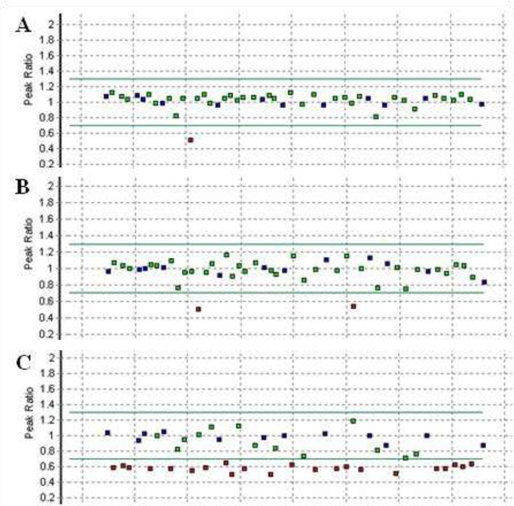
Figure 1.
MLPA findings for 3 reference DNAs which showed abnormal probe levels. A) Individual with a deletion of PMS2 exon 14. B) Individual with an apparent deletion of exons 14 and 15 from PMS2-CL. C) Confirmation that the entire PMS2 coding region is deleted in an individual previously classified as having an exon 1 – 12 deletion with the older version of the MLPA kit. Amounts of specific probes, relative to a set of control samples which have two copies of each probe, are represented by squares. A red square denotes a probe whose copy number is suggestive of a gain or a deletion. Sequencing across the probe binding sites in both PMS2 and PMS2-CL allows for deletions and duplications to be assigned to specific loci. A detailed description of the analyses can be found in Vaughn et al. (2011).1

