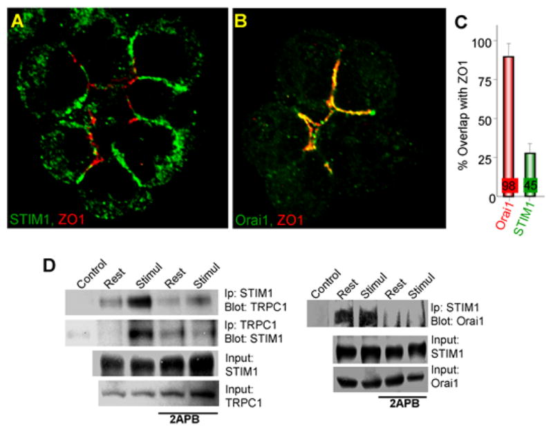
Fig. 2. Localization of NATIVE STIM1 and Orai1 in polarized cells.

In panels (A–C), ZO1 was used to mark the tight junction at the apical pole of mouse pancreatic acinar cells. Panel (A) shows that small fraction of native STIM1 is at the apical pole and most of native STIM1 is at the lateral plasma membrane and panel (B) shows that native Orai1 is confined exclusively to the apical pole. The fraction of native STIM1 and native Orai1 at the apical pole is shown in panel (C). All experiments in panels (A–C) are with store-depleted cells. Panel (D) shows that native STIM1 and TRPC1 and STIM1 and Orai1 are co-immunoprecipitated, the co-IP of STIM1 and TRPC1 (but not of STIM1 and Orai1) is enhanced by cell stimulation that depletes the Ca2+ stores and the complexes are broken with 2APB, which dissociates STIM1-formed complexes. The figure is reproduced from (Hong et al., 2010).
