Figure 1.
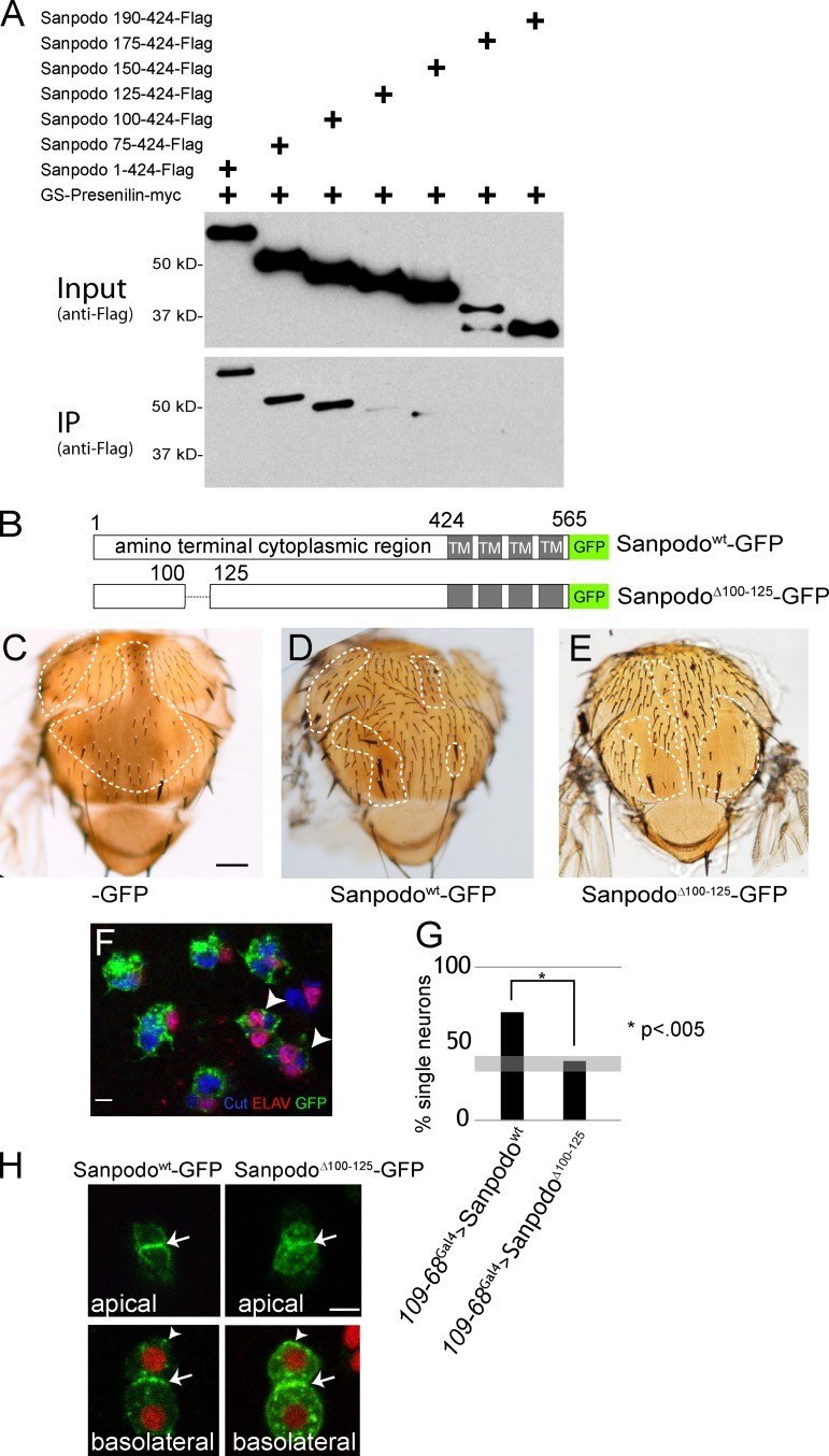
Sanpodo’s interaction with Presenilin is required for pIIa cell fate. Coexpression of the γ-secretase complex with myc-tagged Presenilin and Flag-tagged Sanpodo amino-terminal fragments in Drosophila S2 cells. (A) Binding of the amino-terminal cytoplasmic portion of Sanpodo (amino acids 1–424) to Presenilin-myc requires amino acids 100–125. (B) Schematic of SanpodoΔ100–125 mutant, deleting amino acids 100–125 within the amino terminus. (C–E) Thoraces of flies with sanpodoc55Sb e mutant clones (dotted line: approximate clone boundaries, as determined by e/e and Sb/Sb) with 109-68-Gal4–driven expression of GFP, Sanpodowt-GFP, or SanpodoΔ100–125–GFP using the MARCM system. Sanpodowt-GFP expression largely restores the wild-type bristle pattern to the thorax in sanpodo mutant clones (C; bar, 200 µm), whereas SanpodoΔ100–125–GFP expression fails to suppress the characteristic balding (D). Supernumerary neurons (marked with both Cut [blue] and ELAV [red]) in pupal sanpodoC55 mutant sensory organs expressing SanpodoΔ100–125–GFP (green) using the MARCM system (F, arrowhead). Bar, 5 µm. (G) Quantification of pupal sanpodo mutant sensory organs with only one ELAV+ neuron, indicating suppression of the sanpodo mutant phenotype; when expressing either Sanpodowt-GFP or SanpodoΔ100–125–GFP in MARCM clones using 109-68-Gal4, expression of SanpodoΔ100–125–GFP does not suppress the incompletely penetrant sanpodo mutant phenotype (gray bar indicates previously reported range of single neuron sensory organs in sanpodo null mutant clones; Jafar-Nejad et al., 2005; Roegiers et al., 2005). (H) SanpodoΔ100–125–GFP localizes to the apical and basolateral plasma membrane (white arrows) and intracellular puncta (white arrowheads) in pIIb/pIIa cells, similarly to wild-type Sanpodo-GFP (Histone RFP [red], Sanpodowt-GFP and SanpodoΔ100–125–GFP [green]). Bar, 2 µm.

