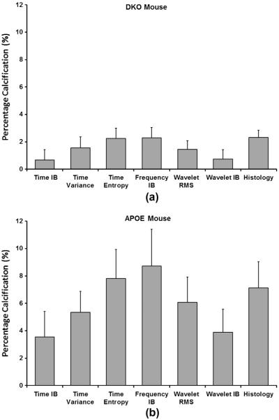
Fig. 5.
Measurements of calcified areas as a percentage of vessel cross-sections evaluated from ultrasound hybrid imaging and histology in atherosclerotic mice. (a) Percentage calcification calculated for a DKO mouse, and (b) percentage calcification calculated for an APOE-KO mouse. Ultrasound parameters include time integrated backscatter (TIB), time variance (Tvar), time entropy (TE), frequency integrated backscatter (FIB), wavelet RMS (Wrms), and wavelet integrated backscatter (WIB), respectively. Cross-sections were acquired at the bifurcation of the aortic arch. Error bars represent the standard error (p < 0.05). Ultrasound parameters were able to detect calcified lesions and some parameters including TE, FIB, and Wrms showed good measurements agreement with histology for percentage calcification (n = 4 cross-sections).

