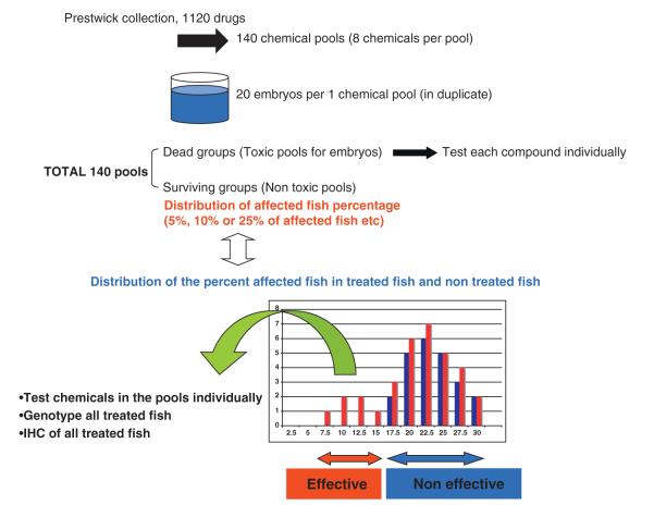
Fig. 1.
Schematic of drug screening using DMD model fish.In the first screen, twenty, one dpf-embryos from a mating of sapje heterozygous fish were arrayed, twenty per well, in 48-well plates containing 0.25 ml of fish water and eight pooled chemicals (vertical pool of eight chemicals in 96 well-plate) from the prestwick collection (total 1120 chemicals) in duplicate. The chemicals were at a final concentration of 2.4 μg/ml. As a normal control, twenty embryos were cultured in duplicate without chemicals. At 4 dpf, the birefringence of all fish was tested using a dissecting microscope. Since the muscle phenotype in these mutant fish is transmitted in a recessive manner, approximately, 25% of the offspring have the abnormal muscle birefringence phenotype after 4 dpf. After comparison of the percentage of affected fish at 4 dpf between chemical-treated fish (red bars) and non-treated fish (blue bars), chemical pools with a reduced percentage of affected fish upon birefringence assay compared to those in non-treated fish were selected for the secondary screen and were tested individually. All surviving fish, both normal and those with apparently restored muscle, were sacrificed. Heads were removed for genotyping and bodies were used for immunostaining.

