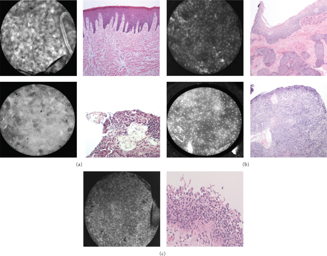
Figure 8.
Current limitations of the HRME. (a) Keratin artifact appears hyperfluorescent, obscuring visualization of the nuclei in benign mucosa from the hard palate (top) and malignant mucosa from the base of tongue (bottom) ( H&E original magnification 100x). (b) Underlying invasive squamous cell carcinoma infiltrating beneath benign squamous epithelium in the larynx (top) and tonsil (bottom) (H&E original magnification 100x). (c) Pseudostratified columnar epithelium of respiratory mucosa from the larynx (H&E original magnification 100x). Notice the relatively crowded nuclei which may be confused with dysplasia or carcinoma.

